- Detects molecules as small as 0.5 kDa.
- Measures binding affinities over a wide dynamic range, including high-affinity interactions in the ~100 pM range.
For more information click here

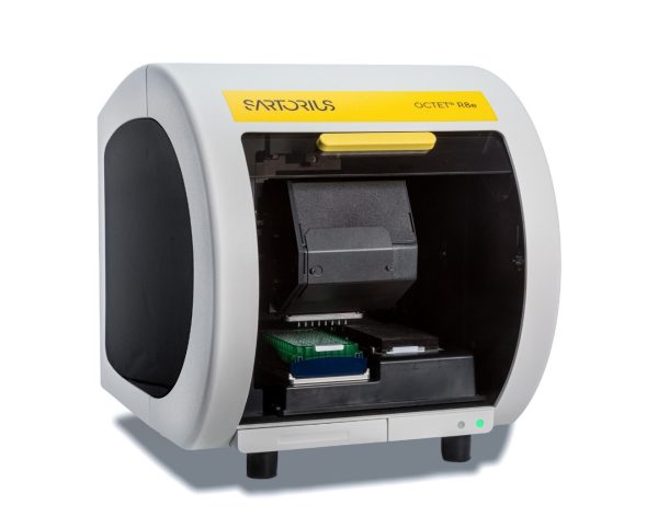
The Octet R8e is a BLI (Bio-Layer Interferometry)-based instrument used to measure the binding affinity between two molecules.
Samples are loaded into 96- or 384-well plates. The ligand is immobilized on an optical sensor, allowing up to eight samples to be analyzed simultaneously. The sensor is then incubated with the analyte, and the interaction is monitored in real time to determine the association rate (Ka), dissociation rate (Kd), and equilibrium dissociation constant (KD).
A wide variety of biosensors is available for capturing His-tagged, biotin-tagged, and Fc-tagged proteins. Alternatively, ligands can be immobilized using standard amine coupling chemistry. Specialized biosensors for nanobody capture are also available.
For more information click here
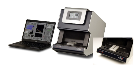
The Monolith NT.115 measures affinity in steady state by using small amounts of samples (ul range) as follows:
• One molecule (that is labeled with a fluorescent blue\green excited dye) is incubated with dilutions of the other molecules.
• The mixed samples are transferred to capillaries and subjected to a temperature gradient.
• Changes in fluorescence (due to heat or migration) are monitored
• A plot of change in fluorescence vs. concentration helps calculate the affinity at steady state.
The whole process can take less than 30 minutes.
For more information, contact Alon Zilka
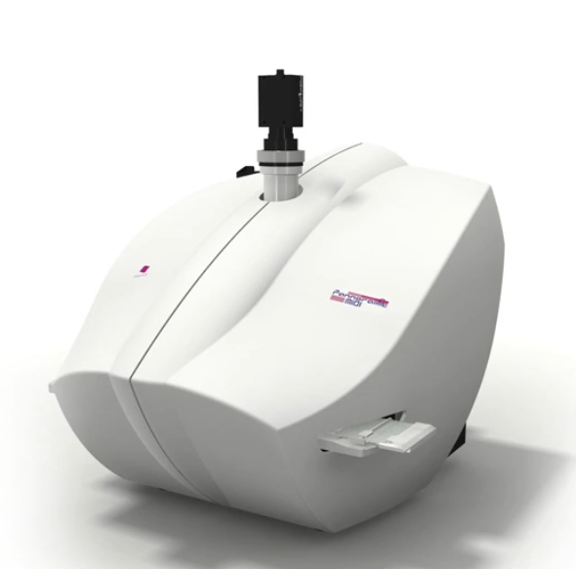
Automated brightfield\fluorescent slide scanner
The midi Panoramic scanner from 3DHISTECH is an automated slide scanner able to scan Histochemical or fluorescent stained slides.
Important:
For optimum performance slides should be up to 1.03 mm thick. Thicker slides are not allowed.
Changing between the Fluorescent and the brightfield camera is done manually so only one type of staining should be run at a time.
The instrument is equipped with an X20 0.8NA and can read 4 fluorescent dyes (DAPI, Cye2, TRITC and Cy5 equivalent dyes) in its fluorescent mode.
The Zeiss Elyra 7 is a state-of-the-art super-resolution imaging platform available at our facility, offering exceptional flexibility for both live-cell and fixed-sample imaging. It combines multiple advanced imaging modalities—including Lattice SIM², Single Molecule Localization Microscopy (SMLM), and Apotome optical sectioning—to provide nanoscale resolution, fast acquisition speeds, and compatibility with a wide range of biological samples.
Lattice SIM²: Fast, Gentle, and Super-Resolved
Lattice SIM² is an advanced structured illumination method that enables super-resolution imaging down to ~60 nm while maintaining low phototoxicity—making it ideal for live-cell experiments. It uses a lattice pattern of light to extract high-resolution information from standard fluorophores and produces fast, high-contrast 3D images with minimal photodamage.
To further enhance performance:
• Leap mode enables rapid volumetric imaging with improved axial resolution, making it ideal for capturing fast 3D biological processes.
• Burst mode captures sequences at extremely high speed to reduce motion blur and allow real-time observation of rapid cellular events.
________________________________________
SMLM (PALM/STORM): Nanoscale Precision, Molecule by Molecule
Single Molecule Localization Microscopy (SMLM) techniques, such as PALM and STORM, push resolution down to 20 nm or better by localizing individual fluorophores with extreme precision. This mode is best suited for fixed samples where fine molecular details—such as protein clusters, membrane domains, or cytoskeletal structures—need to be resolved.
________________________________________
Apotome: Enhanced Widefield Imaging
The Elyra 7 also includes Apotome, a structured illumination technique for widefield microscopy that removes out-of-focus light to produce optically sectioned images. It’s a fast and simple way to improve contrast and resolution in thicker specimens, especially for routine fluorescence imaging.
________________________________________
With these advanced imaging capabilities, the Elyra 7 system supports a broad spectrum of applications—from live-cell dynamics and 3D organelle tracking to ultra-high-resolution structural mapping of fixed samples. Contact us to learn more, schedule training, or reserve time on the system.
Zeiss Celldiscoverer 7 (CD7) is a widefield based platform that combines several hardware and software element to allow long lasting live cell fluorescent and brightfield imaging. The system can accommodate wide verity of sample carriers (glass or plastic) and can be easily used with multi-well plate. CD7 has unique adjustable optical setup that supports wide range of working distances. In combination with Zen image analysis tools, CD7 can generate high-quality and high-throughput data on single cell level (“flow cytometry like”) at spatial and temporal dimensions. CD7 is also suitable platform to image large sample such as tissue sections.
Tutorial: CellDiscoverer 7
Please see description of available software tools at the technical specification sheet .

The Image Analysis service offers:
405, 488, 561 and 640nm excitation lasers
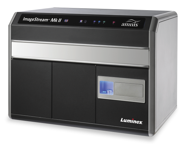
High throughput imaging of cells and particales in liquid suspension. By combining the speed, sensitivity, and phenotyping abilities of flow cytometry with the detailed imagery and functional insight of microscopy, the ImageStreamX Mark II overcomes the limitations of both techniques and opens the door to an extensive range of novel applications.
4 Fluorescent channels, 20x 40x 60 objectives. IDEAS data analysis software.
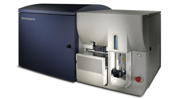
Fluorescence-Activated Cell Sorting (FACS) is a method for analysis and sorting of cells and other biological particles in liquid suspension(e.g. exosomes) based on light scattering and fluorescence characterizations.
4 excitation lasers (405nm, 488nm, 561nm, 635nm). 13 Fluorescent channels. up to 4 populations sorting, Single cell sorting
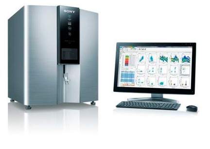
The SP6800 is a FACS spectral analyzer specially adapted to analyze multi stain fluorescently labelled cells without the need for complicated compensation corrections.
By using a configuration of 32 filters between the 500 and the 800nm range, and an additional set of 5 more filters between 400 and 500nm range, it can calculate the emission spectrum of each dye used in the experiment. Next, it calculates its contribution to the total fluorescence. Finally, it easily displays the results in a multicolor staining experiment, without the need for biased\complicated compensation matrixes.
The results are then displayed as a standard density plot/histogram as in any other FACS instrument and can be also exported in a FSC 3.0 file format.
405, 488 and 640 nm excitation lasers
For more information, on the Flowjo site, please click here
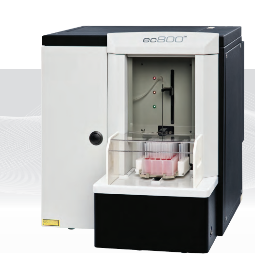
The EC800 can analyze cells based on their Size, density, expression of fluorescent proteins and the ability to bind fluorescent Ab’s.
Has a 488 and 640nm lasers.
Contains an auto loader unit for tubes and plates.