- Zeiss Elyra 7 Super-Resolution Microscopy
- Zeiss Celldiscoverer 7 – Automated live/fixed fluorescence imaging
- Image analysis services
- Support for processing and analyzing fluorescence microscopy images
- Implementation and integration of tools for obtaining quantitative information from micrographs
- Consultation for optimizing output in microscopy, and training of researchers on the use of bioimage analysis tools
- Zeiss LSM880 Airyscan
- Fv1000 laser scanning confocal
- ImageStreamX mkII
- FACSAria III
- SY3200 cell sorter
- SP6800 spectral analyzer
- EC800 cell analyzer
- Spinning Disk
- ProteOn XPR36
- Carboxyl coated chips (for binding proteins via an amine group)
- Neutravidin coated chips (for binding a biotinylated molecule)
- Tris-NTA coated chips (for binding his tagged proteins)
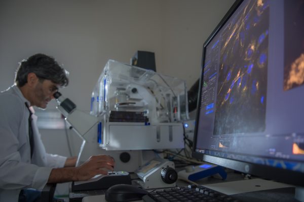
The Zeiss Elyra 7 is a state-of-the-art super-resolution imaging platform available at our facility, offering exceptional flexibility for both live-cell and fixed-sample imaging. It combines multiple advanced imaging modalities—including Lattice SIM², Single Molecule Localization Microscopy (SMLM), and Apotome optical sectioning—to provide nanoscale resolution, fast acquisition speeds, and compatibility with a wide range of biological samples.
________________________________________
Lattice SIM²: Fast, Gentle, and Super-Resolved
Lattice SIM² is an advanced structured illumination method that enables super-resolution imaging down to ~60 nm while maintaining low phototoxicity—making it ideal for live-cell experiments. It uses a lattice pattern of light to extract high-resolution information from standard fluorophores and produces fast, high-contrast 3D images with minimal photodamage.
To further enhance performance:
• Leap mode enables rapid volumetric imaging with improved axial resolution, making it ideal for capturing fast 3D biological processes.
• Burst mode captures sequences at extremely high speed to reduce motion blur and allow real-time observation of rapid cellular events.
________________________________________
SMLM (PALM/STORM): Nanoscale Precision, Molecule by Molecule
Single Molecule Localization Microscopy (SMLM) techniques, such as PALM and STORM, push resolution down to 20 nm or better by localizing individual fluorophores with extreme precision. This mode is best suited for fixed samples where fine molecular details—such as protein clusters, membrane domains, or cytoskeletal structures—need to be resolved.
________________________________________
Apotome: Enhanced Widefield Imaging
The Elyra 7 also includes Apotome, a structured illumination technique for widefield microscopy that removes out-of-focus light to produce optically sectioned images. It’s a fast and simple way to improve contrast and resolution in thicker specimens, especially for routine fluorescence imaging.
________________________________________
With these advanced imaging capabilities, the Elyra 7 system supports a broad spectrum of applications—from live-cell dynamics and 3D organelle tracking to ultra-high-resolution structural mapping of fixed samples. Contact us to learn more, schedule training, or reserve time on the system.
Zeiss Celldiscoverer 7 (CD7) is a widefield based platform that combines several hardware and software element to allow long lasting live cell fluorescent and brightfield imaging. The system can accommodate wide verity of sample carriers (glass or plastic) and can be easily used with multi-well plate. CD7 has unique adjustable optical setup that supports wide range of working distances. In combination with Zen image analysis tools, CD7 can generate high-quality and high-throughput data on single cell level (“flow cytometry like”) at spatial and temporal dimensions. CD7 is also suitable platform to image large sample such as tissue sections.
Please see description of available software tools at the technical specification sheet .
The Image Analysis service offers:
Confocal microscope with super resolution (140nm) and spectral imaging
6 excitation lasers (405nm, 458bm, 488nm, 514nm, 561nm, 633nm). 2 PMT detectors, 32 Channels GaAsp spectral detector, Airyscan imaging. fully incubated for live cell imaging. Wide selection of objectives for different application. 2 excitation lasers (488nm, 642nm).
405, 488, 561 and 640nm excitation lasers
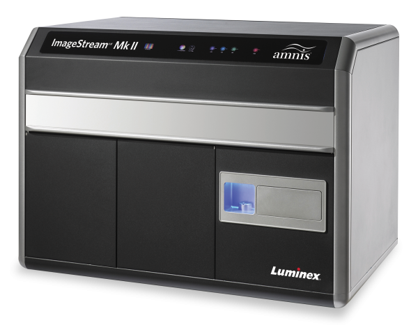
High throughput imaging of cells and particales in liquid suspension. By combining the speed, sensitivity, and phenotyping abilities of flow cytometry with the detailed imagery and functional insight of microscopy, the ImageStreamX Mark II overcomes the limitations of both techniques and opens the door to an extensive range of novel applications.
4 Fluorescent channels, 20x 40x 60 objectives. IDEAS data analysis software.
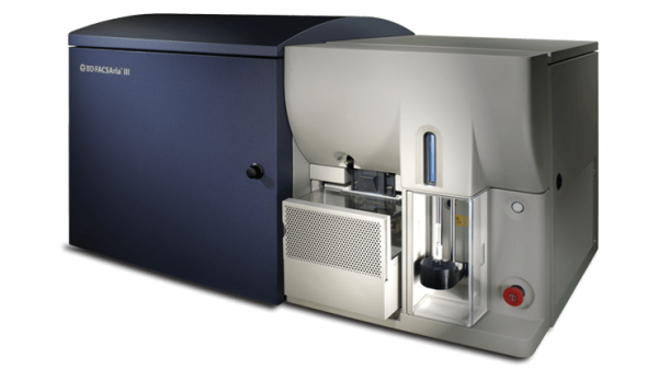
Fluorescence-Activated Cell Sorting (FACS) is a method for analysis and sorting of cells and other biological particles in liquid suspension(e.g. exosomes) based on light scattering and fluorescence characterizations.
4 excitation lasers (405nm, 488nm, 561nm, 635nm). 13 Fluorescent channels. up to 4 populations sorting, Single cell sorting
The SY3200 can analyze\sort cells based on their Size, density, expression of fluorescent proteins and the ability to bind fluorescent Ab’s
2 sorting modules:
HAPS1: 375nm, 488nm, 561nm and 590nmc excitation lasers
HAPS2: 405, 488 and 640nm excitation lasers.
13 Fluorescent channels. up to 4 populations sorting, Single cell sorting
Located in a biological hood for sterile sorting conditions.
The SP6800 is specially adapted to analyze multi stain fluoresenctly labelled cells without the need for complicated compensation corrections
405, 488 and 640 nm excitation lasers
The EC800 can analyze cells based on their Size, density, expression of fluorescent proteins and the ability to bind fluorescent Ab’s
Has a 488 and 640nm lasers.
Contains an auto loader unit for tubes and plates.
Confocal imaging via spinning disk involves scanning a field with laser light from a number of pinholes arranged in a pattern on a modified Nipkow disk. Unlike laser scanning confocal microscopes (LSM) which scan one point of laser light across an entire field, a spinning disk confocal scans approximately 1,000 points of laser light across the field simultaneously resulting in much faster image production. In a traditional LSM, the detector is a photomultiplier tube which can register the signal from only one point of light (pixel) at a time and with a typical quantum efficiency of 40-50%. In an SDC the detector is a CCD camera which can register the signal from a quarter million or million pixels simultaneously with a quantum efficiency upwards of 95%. The result is that while LSMs can typically image on the order of one full frame per second, SDCs can image at over 1,000 frames per second. This significant speed difference combined with the superior sensitivity of high-end CCDs has made spinning disk confocal a must-have technology for advanced live cell imaging labs.
For technical specification, please click here
and for more information, click here
Measures affinity and kinetic parameters between molecules in high throughput mode
6 channels for ligand binding and 6 channels for analyte bindings
Various chips (for ligand binding) can be purchased from the manufacture (Xantec):
Other chips are also available
All chips can be bought with various binding site densities to optimize detection of analyte binding to the ligand.


